Home>Health & Wellness>Common Health Issues>How To Administer Dental X-Rays On Dogs

Common Health Issues
How To Administer Dental X-Rays On Dogs
Published: January 31, 2024
Learn how to administer dental x-rays on dogs to identify common health issues. Our comprehensive guide will help you ensure your pet's dental health.
(Many of the links in this article redirect to a specific reviewed product. Your purchase of these products through affiliate links helps to generate commission for Pawsomeoldies.com, at no extra cost. Learn more)
Table of Contents
Introduction
Dental health is a crucial aspect of a dog's overall well-being. Just like humans, dogs can experience dental issues such as cavities, gum disease, and tooth decay. However, unlike humans, dogs cannot communicate their dental discomfort, making it challenging for pet owners to identify potential problems. This is where dental x-rays play a vital role in ensuring the oral health of our furry companions.
Dental x-rays, also known as dental radiographs, provide a comprehensive view of a dog's oral cavity, including the teeth, roots, and surrounding bone structure. They are instrumental in diagnosing hidden dental problems that may not be apparent during a routine oral examination. By revealing issues beneath the gum line, dental x-rays enable veterinarians to accurately assess the extent of dental disease and formulate an effective treatment plan.
In this article, we will delve into the process of administering dental x-rays on dogs, shedding light on the significance of this diagnostic tool and the steps involved in obtaining and interpreting the x-ray images. Understanding the importance of dental x-rays and the techniques used to capture and analyze these images is essential for both veterinary professionals and pet owners. By gaining insight into this aspect of canine healthcare, individuals can actively contribute to the well-being of their canine companions and ensure that any dental issues are promptly identified and addressed.
As we embark on this exploration of dental x-rays for dogs, it's important to recognize the impact of proactive dental care on a dog's quality of life. By proactively addressing dental problems through the use of x-rays and subsequent treatments, we can help our canine friends maintain healthy teeth and gums, alleviate discomfort, and prevent more serious health complications. With this understanding, let's delve into the world of dental x-rays for dogs, uncovering the valuable role they play in safeguarding the oral health of our beloved pets.
Understanding the importance of dental x-rays for dogs
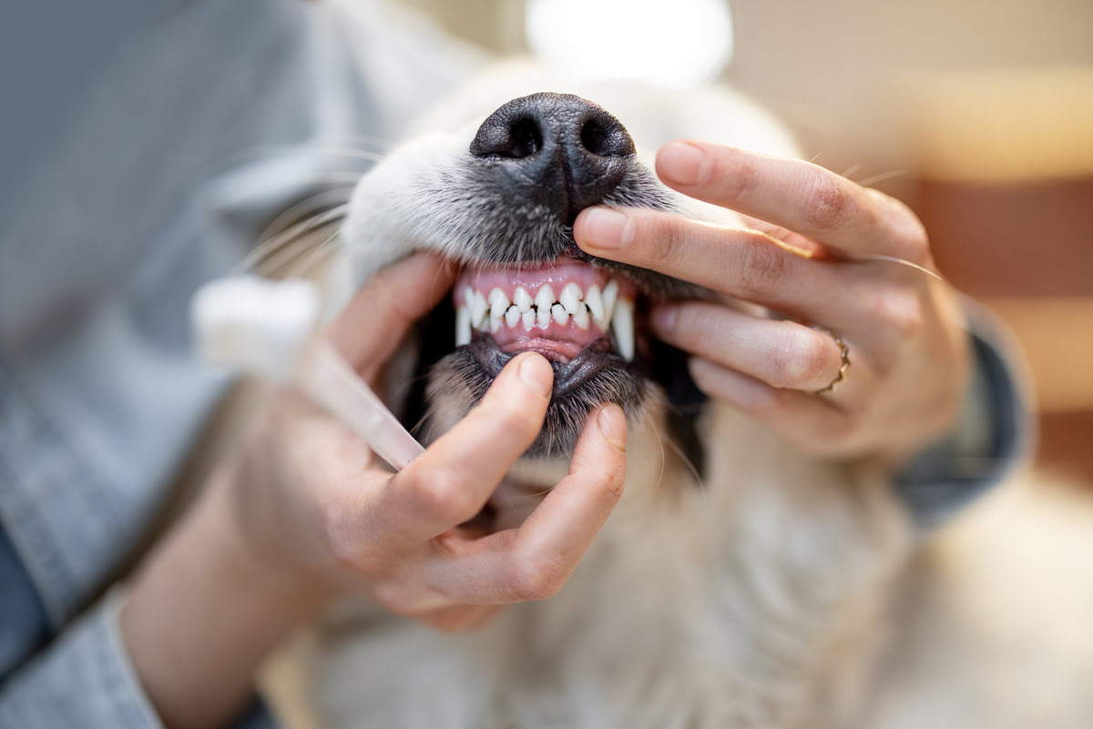
Dental x-rays are an indispensable tool in the realm of veterinary dentistry, offering a comprehensive view of a dog's oral health that extends beyond what is visible to the naked eye. While a visual examination of a dog's teeth and gums provides valuable insights, it often falls short in detecting underlying dental issues that may be lurking beneath the surface. This is where dental x-rays step in, playing a pivotal role in uncovering hidden problems and guiding effective treatment strategies.
One of the primary reasons dental x-rays are crucial for dogs is their ability to reveal dental problems that are not apparent during a routine oral examination. Dogs, like humans, can experience a range of dental issues, including periodontal disease, tooth fractures, abscesses, and root abnormalities. However, these problems may not manifest visibly, especially in the early stages. Dental x-rays enable veterinarians to peer beneath the gum line, where many dental issues originate, and identify abnormalities that require attention.
Moreover, dental x-rays aid in assessing the overall dental health of dogs by providing a detailed view of the teeth, roots, and surrounding bone structure. This comprehensive perspective allows veterinarians to evaluate the extent of dental disease, determine the presence of hidden lesions or tumors, and make informed decisions regarding treatment options. By gaining a thorough understanding of the dog's oral health through x-ray imaging, veterinarians can tailor treatment plans to address specific issues and alleviate any discomfort or pain experienced by the dog.
Furthermore, dental x-rays are instrumental in monitoring the progression of dental conditions and evaluating the success of treatments. By comparing current x-ray images with previous ones, veterinarians can assess changes in the dog's dental health, track the effectiveness of interventions, and make adjustments to treatment plans as needed. This proactive approach to dental care enables early detection of potential complications and ensures that appropriate measures are taken to maintain the dog's oral well-being.
In essence, the importance of dental x-rays for dogs lies in their ability to uncover hidden dental problems, provide a comprehensive view of oral health, guide tailored treatment plans, and facilitate ongoing monitoring of dental conditions. By harnessing the insights offered by dental x-rays, veterinarians and pet owners can work together to safeguard the dental health of dogs, promoting overall well-being and enhancing their quality of life.
Preparing for dental x-rays
Before embarking on the process of obtaining dental x-rays for a dog, thorough preparation is essential to ensure a smooth and effective procedure. The preparation phase encompasses several key elements that contribute to the successful acquisition of high-quality x-ray images while prioritizing the safety and well-being of the dog.
Ensuring a Calm and Comfortable Environment
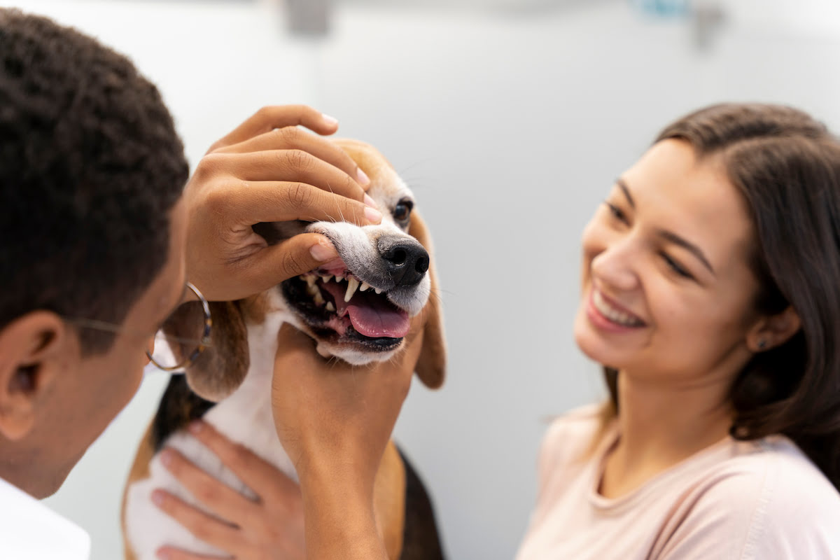
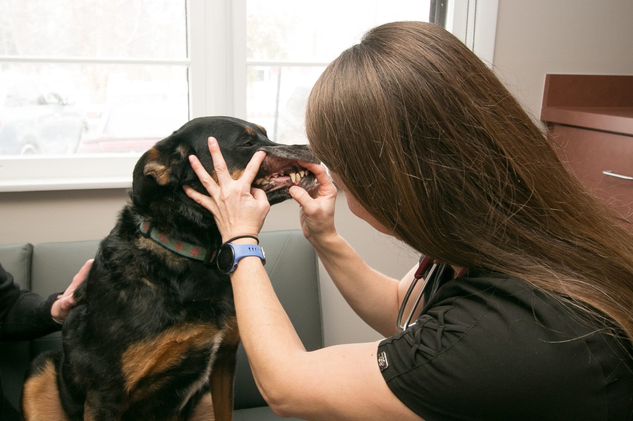
Creating a calm and comfortable environment is paramount when preparing for dental x-rays. Dogs are highly sensitive to their surroundings, and a relaxed atmosphere can significantly reduce their anxiety and stress during the procedure. This involves selecting a quiet and familiar space within the veterinary clinic where the dog can feel at ease. Minimizing loud noises and disturbances and providing comforting measures, such as soft bedding and familiar scents, can help alleviate the dog's apprehension and promote a sense of security.
Securing Restraint and Safety Measures
Proper restraint and safety measures are crucial components of preparing for dental x-rays. Ensuring the dog is safely and securely positioned for the procedure is essential for obtaining accurate and clear x-ray images. This may involve the use of gentle restraint techniques, such as specialized harnesses or gentle handling, to keep the dog in the optimal position for the x-ray equipment. Additionally, protective measures, including lead aprons and thyroid collars, should be employed to shield both the dog and veterinary staff from unnecessary radiation exposure.
Read more: Who Performs Dental Prophylaxis In Dogs
Collaborating with Veterinary Professionals
Collaboration with experienced veterinary professionals is fundamental in preparing for dental x-rays. Veterinary technicians and radiology specialists play a pivotal role in guiding the preparation process, offering expertise in positioning the dog for x-ray imaging and ensuring adherence to safety protocols. Their knowledge and proficiency contribute to the seamless execution of the x-ray procedure, fostering a sense of confidence and reassurance for both the dog and their human companions.
Conducting Pre-X-ray Assessments
Conducting pre-x-ray assessments is an integral part of the preparation phase, allowing veterinarians to evaluate the dog's overall health and identify any potential contraindications to the x-ray procedure. This may involve a comprehensive physical examination to assess the dog's fitness for anesthesia, if required, and to address any underlying health concerns that may impact the x-ray process. By conducting thorough pre-x-ray assessments, veterinarians can mitigate potential risks and tailor the procedure to the specific needs of the dog.
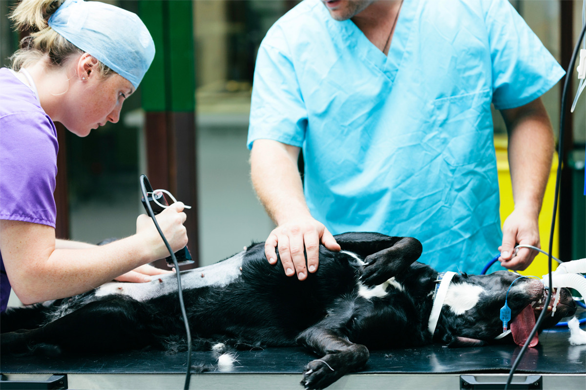
Implementing Anesthesia Protocols (if necessary)
In cases where dogs exhibit significant discomfort or require extensive dental imaging, the implementation of anesthesia protocols may be necessary to ensure their safety and well-being during the x-ray procedure. Anesthesia allows for precise positioning and immobilization, minimizing movement artifacts and enhancing the quality of the x-ray images. Prior to administering anesthesia, comprehensive evaluations of the dog's health status and anesthesia tolerance are conducted to mitigate potential risks and optimize the anesthesia protocol for the individual dog.
In essence, the preparation for dental x-rays encompasses creating a calming environment, ensuring proper restraint and safety measures, collaborating with veterinary professionals, conducting pre-x-ray assessments, and implementing anesthesia protocols when necessary. By meticulously addressing these preparatory elements, veterinary teams can set the stage for a successful and safe dental x-ray procedure, ultimately contributing to the comprehensive assessment of the dog's oral health and the formulation of targeted treatment plans.
Positioning the dog for x-rays
Positioning the dog for dental x-rays is a critical aspect of the imaging process, as it directly influences the quality and accuracy of the resulting x-ray images. Proper positioning not only ensures comprehensive coverage of the dog's oral cavity but also minimizes the need for retakes, reducing the dog's exposure to radiation and streamlining the overall procedure.
The positioning of the dog for dental x-rays involves several key considerations to optimize the imaging process and facilitate the acquisition of clear and detailed x-ray images. Veterinary professionals employ specific techniques to position the dog effectively, taking into account factors such as the type of x-ray being performed, the dog's size and temperament, and the areas of interest within the oral cavity.
One fundamental aspect of positioning the dog for x-rays is the use of gentle restraint to maintain the dog in the desired position for imaging. This may involve the assistance of veterinary technicians or specialized positioning aids to ensure that the dog remains still and cooperative throughout the process. By employing gentle and reassuring handling techniques, veterinary professionals can minimize the dog's anxiety and promote a sense of security, contributing to a more relaxed and cooperative demeanor during positioning.
Additionally, the positioning of the x-ray equipment plays a crucial role in capturing comprehensive images of the dog's oral structures. Veterinary professionals carefully align the x-ray machine to target specific areas of the oral cavity, adjusting the equipment to achieve optimal angles and coverage. This meticulous approach allows for the precise capture of dental structures, including the teeth, roots, and surrounding bone, facilitating a thorough assessment of the dog's dental health.
Furthermore, the use of positioning aids, such as foam blocks or bite-wing holders, assists in stabilizing the dog's head and maintaining consistent positioning during the x-ray procedure. These aids help minimize movement artifacts and ensure that the resulting images accurately reflect the dog's dental anatomy, enabling veterinarians to make precise diagnoses and formulate targeted treatment plans.
In cases where dogs require sedation or anesthesia for the x-ray procedure, positioning becomes even more crucial to ensure the safety and well-being of the sedated animal. Veterinary professionals meticulously position the sedated dog to minimize the risk of airway obstruction and optimize the imaging process while prioritizing the dog's comfort and stability.
In essence, the positioning of the dog for dental x-rays involves a strategic approach that encompasses gentle restraint, precise alignment of x-ray equipment, and the use of positioning aids to facilitate comprehensive imaging. By adhering to these principles, veterinary professionals can ensure that the dog's oral structures are accurately captured, laying the foundation for precise diagnosis and targeted treatment of dental conditions.
Read more: How To Maintain Good Dental Hygiene In Dogs
Taking and processing the x-rays
Taking and processing dental x-rays for dogs involves a meticulous and systematic approach to ensure the acquisition of high-quality images that provide valuable insights into the dog's oral health. This process encompasses several key steps, each playing a crucial role in capturing and interpreting the x-ray images effectively.
Positioning and Exposure
Once the dog is appropriately positioned for the x-ray procedure, the veterinary team carefully adjusts the settings on the x-ray machine to determine the exposure parameters. Factors such as the type of x-ray being performed, the size of the dog, and the specific areas of interest within the oral cavity are taken into consideration to optimize the exposure settings. This meticulous adjustment ensures that the resulting x-ray images offer clear and detailed views of the dental structures, enabling accurate diagnosis and treatment planning.
Image Capture
With the positioning and exposure parameters set, the veterinary professional initiates the image capture process, directing the x-ray machine to emit radiation and capture the desired views of the dog's oral cavity. The dog remains still and cooperative, aided by gentle restraint and calming techniques, to minimize movement artifacts and ensure the clarity of the resulting images. Multiple x-ray views may be taken to comprehensively cover the entire oral cavity, including different angles and perspectives to capture the teeth, roots, and surrounding structures.
Processing and Interpretation
Following the image capture, the x-ray images are processed using specialized equipment to develop and enhance the visibility of dental structures. This processing stage involves the careful handling of the x-ray films or digital images to reveal detailed views of the dog's teeth, roots, and surrounding bone. Veterinary professionals meticulously examine the processed x-ray images, analyzing the dental structures for signs of abnormalities, such as tooth decay, periodontal disease, fractures, or other dental conditions.
Read more: How To Treat A Dental Infection In A Dog
Quality Assurance
Quality assurance measures are integral to the process of taking and processing dental x-rays for dogs. Veterinary professionals meticulously review the developed x-ray images, ensuring that they meet the standards for clarity, detail, and diagnostic value. Any discrepancies or issues with the images are addressed promptly, and additional x-rays may be taken if necessary to obtain comprehensive and accurate views of the dog's oral health.
Documentation and Archiving
Once the x-ray images are processed and deemed satisfactory, they are documented and archived as part of the dog's medical records. These images serve as valuable diagnostic tools for future reference, enabling veterinarians to track the dog's dental health over time, monitor the progression of dental conditions, and assess the effectiveness of treatments. The careful documentation and archiving of x-ray images contribute to the continuity of care and the comprehensive management of the dog's oral well-being.
In essence, the process of taking and processing dental x-rays for dogs involves precise positioning and exposure, meticulous image capture, thorough processing and interpretation, quality assurance measures, and the documentation and archiving of x-ray images. By adhering to these essential steps, veterinary professionals ensure the acquisition of high-quality x-ray images that are instrumental in diagnosing and addressing dental issues, ultimately contributing to the optimal oral health of canine companions.
Interpreting the x-ray results
Interpreting the x-ray results is a pivotal stage in the process of administering dental x-rays for dogs, as it involves analyzing the captured images to glean valuable insights into the dog's oral health. Veterinary professionals employ a systematic approach to interpret the x-ray results, leveraging their expertise to identify potential abnormalities, assess the overall dental condition, and formulate targeted treatment plans.
One of the primary aspects of interpreting x-ray results involves a comprehensive evaluation of the dental structures depicted in the images. This includes scrutinizing the teeth, roots, surrounding bone, and soft tissues for signs of pathology or irregularities. Veterinary professionals meticulously examine the x-ray images, looking for indicators of periodontal disease, tooth decay, fractures, abscesses, and other dental conditions that may impact the dog's oral well-being.
Moreover, the interpretation of x-ray results extends beyond the identification of specific dental issues to encompass an assessment of the overall dental health and the impact of any observed abnormalities. Veterinary professionals analyze the extent and severity of dental conditions, determining the potential implications for the dog's comfort, functionality, and long-term oral health. This comprehensive assessment guides the formulation of tailored treatment plans that address the specific needs and challenges identified through the x-ray interpretation.
Furthermore, interpreting x-ray results involves comparing current images with previous x-rays, when available, to track the progression of dental conditions and evaluate the effectiveness of previous treatments. This comparative analysis enables veterinary professionals to discern changes in the dog's oral health, identify emerging issues, and adjust treatment strategies accordingly. By leveraging the insights gleaned from x-ray comparisons, veterinarians can proactively address evolving dental concerns and optimize the management of the dog's oral well-being.
In essence, interpreting the x-ray results for dogs entails a meticulous examination of dental structures, a comprehensive assessment of dental health, and a comparative analysis of current and previous x-ray images. By harnessing the diagnostic value of x-ray interpretation, veterinary professionals can gain a profound understanding of the dog's oral health, devise targeted treatment approaches, and contribute to the preservation of optimal dental well-being for canine companions.
Conclusion
In conclusion, the administration of dental x-rays for dogs represents a critical component of comprehensive veterinary dental care, offering invaluable insights into the oral health of our canine companions. Through the utilization of advanced imaging techniques and meticulous interpretation of x-ray results, veterinary professionals can effectively diagnose and address a wide array of dental conditions, ultimately contributing to the well-being and comfort of dogs.
The significance of dental x-rays lies in their ability to uncover hidden dental problems that may elude visual examination, providing a comprehensive view of the teeth, roots, and surrounding structures. By leveraging the diagnostic capabilities of dental x-rays, veterinarians can identify issues such as periodontal disease, tooth fractures, abscesses, and root abnormalities, enabling targeted treatment plans tailored to the specific needs of each dog.
Furthermore, the process of preparing for, positioning, capturing, and interpreting dental x-rays involves a meticulous and systematic approach, prioritizing the safety, comfort, and diagnostic accuracy for the dogs. From creating a calm environment and ensuring proper restraint to processing and interpreting x-ray images with precision, each step contributes to the successful acquisition of high-quality x-ray results that guide effective treatment strategies.
The proactive use of dental x-rays facilitates the early detection and management of dental conditions, mitigating potential discomfort and preventing the progression of oral health issues. Additionally, the documentation and archiving of x-ray images enable ongoing monitoring of the dog's dental well-being, supporting continuity of care and informed decision-making for future interventions.
Ultimately, the integration of dental x-rays into canine healthcare exemplifies a commitment to proactive and personalized dental care, aligning with the overarching goal of promoting the overall health and quality of life for our beloved canine companions. By recognizing the pivotal role of dental x-rays and embracing their diagnostic potential, veterinary professionals and pet owners alike can collaborate in safeguarding the oral health of dogs, ensuring that they enjoy a lifetime of healthy smiles and pain-free dental experiences.